Angioma Venoso Cerebral Radiopaedia
Seven patients with angiographically proved cerebral venous angiomas were imaged by magnetic resonance The venous angioma was identified in all cases The most characteristic feature was an enlarged transcerebral draining vein (86%), followed by increased signal on T2weighted images (57%) and decreased signal on T1weighted images (40%) in adjacent parenchyma.

Angioma venoso cerebral radiopaedia. Exploración TAC de tórax La tomografía computarizada (TC) del tórax utiliza un equipo especial de rayos X para examinar anormalidades encontradas en otros exámenes por imágenes, y para ayudar a diagnosticar la causa de una tos sin explicación, la falta de aliento, el dolor de pecho, la fiebre y otros síntomas del pecho. Learning to Live with Brain Injury The Three Rivers Angioma Alliance Community invites you to join author and Angioma Alliance member Deb Brandon as Deb shares with us about her personal experience living with a brain injury We plan to have a question and answer period as time allows When Jan 17, 21 0100 PM Eastern Time (US and Canada. Cavernous angiomas represent approximately 1% of intracranial vascular lesions and 15% of cerebrovascular malformations With the advent of MRI, cavernous angiomas are currently the most commonly.
The MRtechniques that are used for the diagnosis of cerebral venous thrombosis are Timeofflight (TOF), phasecontrast angiography (PCA) and contrastenhanced MRvenography TimeofFlight angiography is based on the phenomenon of flowrelated enhancement of spins entering into an imaging slice. 4 Cerebral 5 Reacciones a drogas 6 Sueros excesivos 7 Embolismo graso 8 Gases tóxicos 9 Heroína 10 4,6Embolismo pulmonar 11 Uremia 12 Trauma torácico RARAS 1 Reacciones alérgicas 2 Colagenosis 3 Ahorcamiento y Sofocación 4 Altitud elevada 5 Hipoproteinemia 6 Malaria 7 Inmersión 8 Toxicidad por oxígeno 9 Neumotórax con. Información precisa y actualizada para pacientes sobre desvío portosistémico intrahepático transyugular Averigüe qué puede sentir, cómo prepararse para el examen, cuáles son los beneficios y los riesgos, y más.
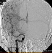
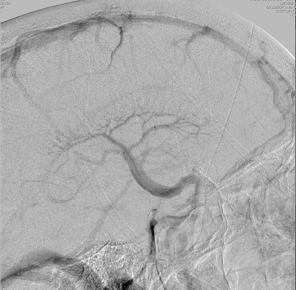
Origen fetal de la arteria cerebral posterior se trata de una variante que se encuentra presente cuando no se produce la regresión de la arteria cerebral posterior en su trayecto fetal Puede observarse una arteria comunicante posterior muy prominente, que aporta la mayor parte del flujo vascular a la arteria cerebral posterior desde la. Cerebral angiomas are vascular abnormalities comprised of clusters of abnormally dilated blood vessels They can be singular or multiple, and are found in the brain, spinal cord, and rarely, in other areas of the body including the skin and retina They are also known as cavernous angioma cavernous hemangioma cavernous venous malformation. Several unusual cases of cerebral venous angiomas as well as some characteristic cases are reported The characteristic angiographic feature is that of a collection of dilated medullary veins draining into a single large draining vein, which appears first in the early venous phase and persists into the late venous phase of the arteriogram.
A developmental venous anomaly (DVA) is an unusual or irregular arrangement of small veins that may look like the spokes of a wheel The veins drain into a larger central vein DVAs are benign (not cancerous) DVAs also may be called venous angiomas or benign. Un angioma cerebral (también denominado hemangioma cerebral) es un tipo de malformación vascular caracterizada por agrupaciones de capilares dilatados Un angioma es una malformación o neoplasia de las células de los vasos sanguíneos que puede aparecer en forma de nódulo o de caverna formada por endotelio que contienen sangre. The venous malformations (or venous angiomas) are developmental anomalies that grow gradually and are considered benign Although aberrant in structure, they are still functional and fullyintegrated with the venous system;.
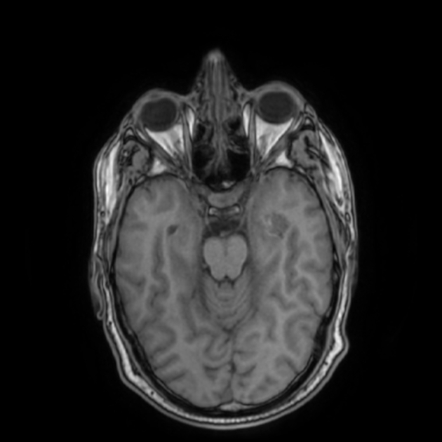
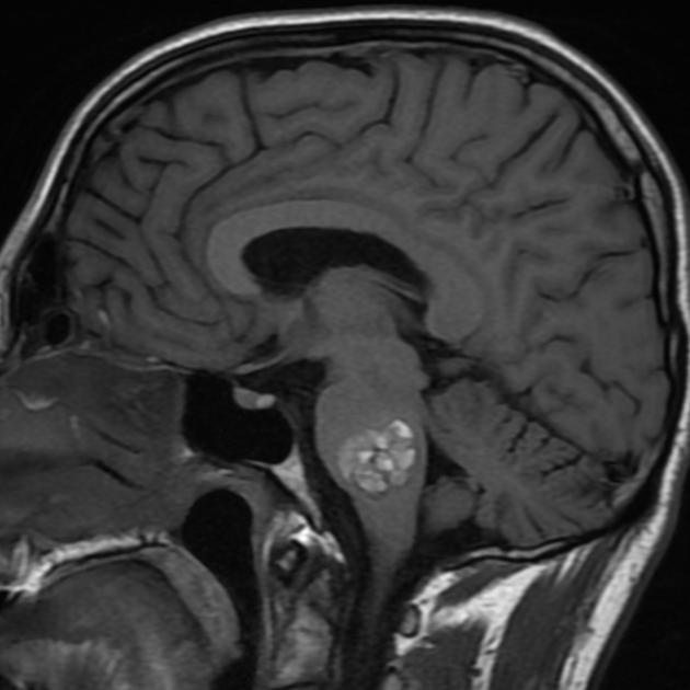
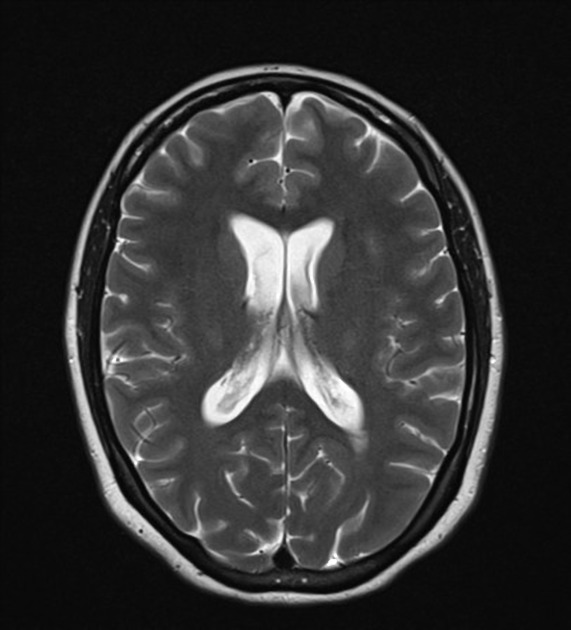
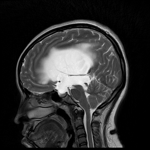
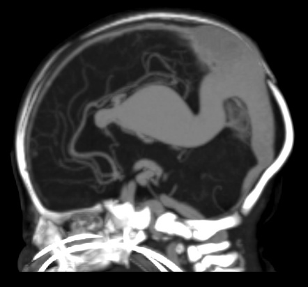
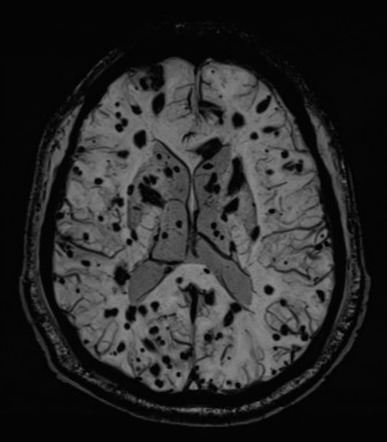
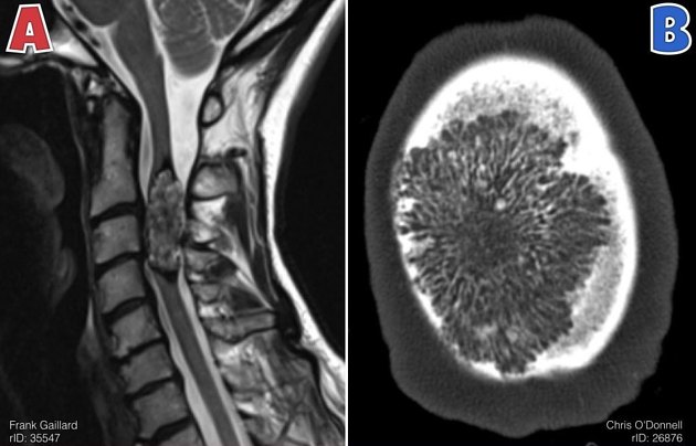
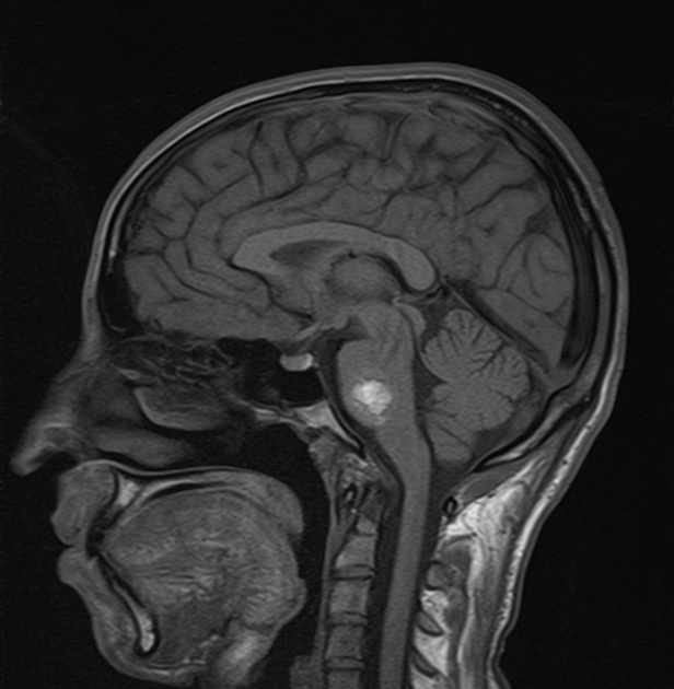
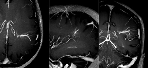
Axial susceptibility images showing tangle of blood vessels with a prominent cortical draining vein and small collector vein towards the frontal horn draining into septal vein typical of a developmental venous anomaly (DVA) also referred to as a venous angioma. Cerebellar Venous Angiomas A Continuing Controversy From the Departments of Neurology (Drs Biller and Toffol), Neurosurgery (Dr Shea), and Radiology (Drs Fine and AzarKia), Loyola University Medical Center, Maywood, Ill Dr Biller is now with the University of Iowa Hospitals and Clinics, Iowa City. Venous angiomas are vascular malformations diagnosed with increasing frequency in the last few years, due to the availability of new neuroimaging techniqueso In this paper we describe a case of venous angioma studied with cerebral angiography, CT and MRI and the literature in relation with this specific vascular lesiono.
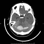
Cavernous angiomas represent approximately 1% of intracranial vascular lesions and 15% of cerebrovascular malformations With the advent of MRI, cavernous angiomas are currently the most commonly. The basal ganglia are a group of grey matter nuclei in the deep aspects of the brain that is interconnected with the cerebral cortex, thalami and brainstem In a strict anatomical sense, it contains three paired nuclei that together comprise the corpus striatum caudate nucleus;. To determine the venous angioma of the brain, radiography is rarely used due to the inability to penetrate the cranium But this method, for the diagnosis, is still acceptable in some cases This analysis is necessary for head trauma or suspicion of it Ultrasonography This method is acceptable when a suspected disease in a newborn baby.
1 Chennai, Tamil Nadu/IN, 2 Chennai/IN. A cavernous angioma is a congenital disorder identified by “a complex, tangled web of arteries and veins” with a short circuit and high pressure caused by arterial blood flowing rapidly in the veins It is also called a cerebral cavernous venous malformation, arteriovenous malformation (AVM), cavernous haemangioma, or cavernoma. The patient in this case is a 32yearold man who presented with a clinical history of headache followed by a worsening of his neurological status Neuroimaging studies demonstrated a brain infarct in the posterior fossa, which was related to thrombosis of the draining vein of a cerebral venous angioma.
ECR / C MR Spectroscopy in paediatric brain lesions – A problem solving tool A Damodaran Nampoothiry 1, K Gopinathan 2, P D J Devimeenal 2, D K GEETHA 2, D R Rajakumaran 2;. Si la MAV ocurre en el cerebro, el sangrado puede causar un derrame o daño cerebral Las MAVs periféricas se desarrollan afuera de la cabeza, del cuello, o de la columna vertebral Reducen el flujo sanguíneo hacia los tejidos circundantes Con el tiempo, esto puede dañar el tejido y causar dolor y úlceras, o llagas, en la piel. Cavernous hemangioma, also called cavernous angioma, cavernoma, or cerebral cavernoma is a type of benign vascular tumor or hemangioma, where a collection of dilated blood vessels form a lesion The abnormal tissue causes a slowing of blood flow through the cavities, or "caverns" The blood vessels do not form the necessary junctions with surrounding cells, and the structural support from the smooth muscle is hindered, causing leakage into the surrounding tissue It is the leakage of blood, refe.
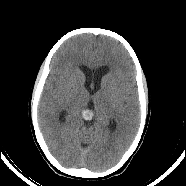
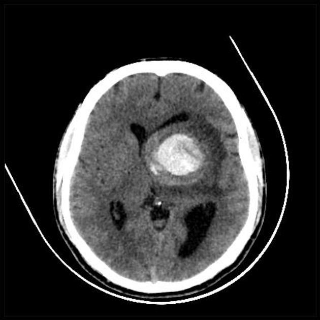
TC cerebral sistema venoso derecho con marcado aumento de calibre RM y Angio RM imagen de señal heterogénea a nivel del lóbulo occipital derecho compatible con angioma cavernoso Imágenes de vasos patológicos de flujo lento, con drenaje venoso anómalo hacia seno lateral derecho. In most cases, Venous Malformations of the Brain arises sporadically, with spontaneous mutation(s) in the TIE2 gene. The MRtechniques that are used for the diagnosis of cerebral venous thrombosis are Timeofflight (TOF), phasecontrast angiography (PCA) and contrastenhanced MRvenography TimeofFlight angiography is based on the phenomenon of flowrelated enhancement of spins entering into an imaging slice.
What are developmental venous anomalies?. Although leptomeningeal angioma is the hallmark of SturgeWeber syndrome (SWS), 1 some patients with suspected SWS show no MRI evidence of pial contrast enhancement 2 A girl with a left port wine stain developed rightsided motor seizures MRI was performed at 18 months of age Postgadolinium T1weighted images (slice thickness 2 mm) showed transmedullary enhancement in the left central. It offers superior delineation of the disease, which is necessary for treatment planning.
The radiological literature on the subject is reviewed and a new classification of cerebral venous angioma based on its pattern of drainage is proposed It is concluded that with the use of thin slices and coronal cuts both the angioma and its pattern of venous drainage can be indentified on CT in a high proportion of cases In addition, with dynamic CT the specificity of CT in diagnosing cerebral venous angioma may further increase. CT of Venous Angiomas of the Brain Preston R Lotz 1 and Ronald G Quisling2 Venous angiomas are very uncommon vascular malfor mations of the brain, which traditionally are described as having no arterial component 1 Recently, however, both their rarity 2 and their lack of enlarged arterial feeders 3 have been questioned The angiographic appearance of venous angiomas has been well described and usually is quite specific 48. Un hemangioma se puede presentar con otras afecciones poco frecuentes Se pueden realizar otros exámenes para buscar problemas relacionados Tratamiento La mayoría de los hemangiomas pequeños o sin complicaciones puede no necesitar tratamiento Con frecuencia desaparecen por sí mismos y la apariencia de la piel regresa a la normalidad.
Venous angiomas are vascular malformations diagnosed with increasing frequency in the last few years, due to the availability of new neuroimaging techniqueso In this paper we describe a case of venous angioma studied with cerebral angiography, CT and MRI and the literature in relation with this specific vascular lesiono. TC cerebral sistema venoso derecho con marcado aumento de calibre RM y Angio RM imagen de señal heterogénea a nivel del lóbulo occipital derecho compatible con angioma cavernoso Imágenes de vasos patológicos de flujo lento, con drenaje venoso anómalo hacia seno lateral derecho. An unusual location for a cavernoma, but these can sometimes be exophytic and extend into the ventricles 1 Note the absence of a hypointense rim on T2 of the ventricular segment, this is known to occur with cavernomas In addition, the character.
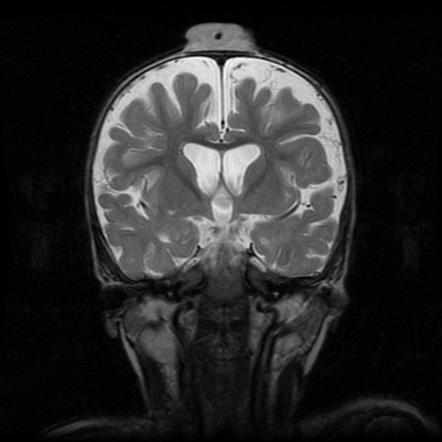
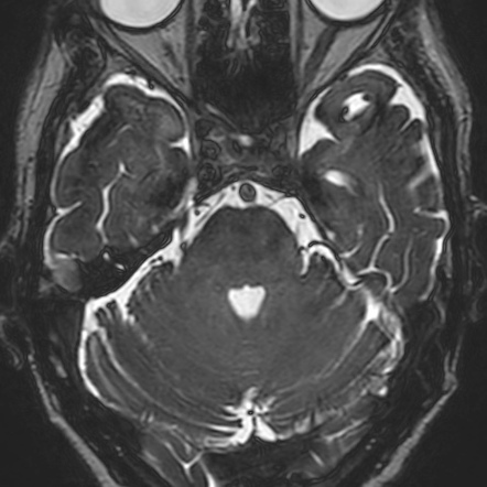
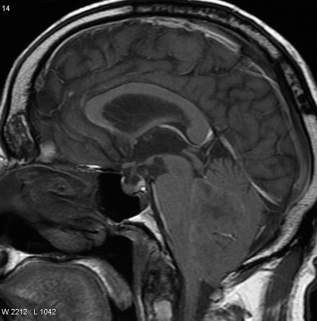
Developmental venous anomaly Developmental venous anomaly in the cerebellum seen on axial contrastenhanced T1 weighted MRI A developmental venous anomaly ( DVA, formerly known as venous angioma) is a congenital variant of the cerebral venous drainage On imaging it is seen as a number of small deep parenchymal veins converging toward a larger collecting vein. Cerebral cavernous venous malformations, also commonly known as cavernous hemangiomas or cavernomas , are common cerebral vascular malformations , usually with characteristic appearances on MRI It is the third most common cerebral vascular malformation after developmental venous anomaly and capillary telangeictasia. Como consecuencia, el angioma venoso se consideraría una causa de la lesión cerebral Otra complicación relacionada con los angiomas venosos es una trombosis de la vena de drenaje Esto puede provocar un infarto venoso, aunque es poco habitual Además, hay estudios que señalan la relación entre el angioma venoso y la malformación cavernosa.
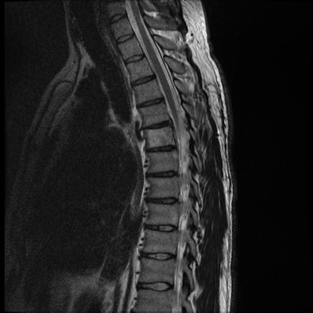
Hemangioma of the spine is one of the most frequent vascular tumors of the skeletal system According to statistics, it suffers every tenth inhabitant of the Earth Among the patients are women and the average age of onset is 30 years It is believed that up to 80% of the fair sex after the age. Cerebral venous angioma (developmental venous anomaly) is the most common cerebral vascular malformation It consists of an intraparenchymal tangle/cluster of dilated medullary veins converging on a single enlarged draining vein It rarely bleeds Differential diagnosis vascular malformation , venous varix. An unusual location for a cavernoma, but these can sometimes be exophytic and extend into the ventricles 1 Note the absence of a hypointense rim on T2 of the ventricular segment, this is known to occur with cavernomas In addition, the character.
El angioma senil también se denomina "en guinda", es de color rojo brillante y suele presentarse en el tronco más que en otra partes del cuerpo Es frecuente en personas mayores de 45 años 2. The basal ganglia are a group of grey matter nuclei in the deep aspects of the brain that is interconnected with the cerebral cortex, thalami and brainstem In a strict anatomical sense, it contains three paired nuclei that together comprise the corpus striatum caudate nucleus;. Brain Tumor, Brain Tumors, Brainstem Neoplasms, Carotid Artery Occlusive Disease, Carotid Stenosis, Cavernous Angioma, Cerebral 1000 Central St Suite 0 Suite 0 Evanston, IL 601 fax Get Directions This location is wheelchair.
Venous angioma of the right hemisphere If we are talking about the hemisphere a layer of gray matter with a thickness of 1345 mm, located on the periphery of the cerebral hemispheres, then the venous angioma of the right hemisphere is fraught with the appearance of such negative symptoms The patient loses the ability to flow smoothly. The cerebrum – Latin for “brain” – is the coordinating center of sensation, intellectual and nervous activity A cavernous malformation is also called a cavernoma or a cerebral cavernous malformation (CCM), but these names are all interchangeable and refer to the same diagnosis Synonyms that refer to the same diagnosis include Cavernoma. What are developmental venous anomalies?.
La anomalía del desarrollo venoso mientras que el cavernoma permanece sin realce hormonales, causas genéticas, siembra a lo largo de un trade biopsia y las anomalías del desarrollo venoso7 La asociación del cavernoma con las anomalías del desarrollo venoso hay que buscarla, pues su frecuencia puede estar. Diagnosis Calcifications can be seen in the radiological examinations due to phleboliths (calcified thrombi) within venous hemangiomas MRI is the imaging modality of choice for diagnosing venous hemangioma;. La hemorragia intracerebral puede deberse a un trauma (lesión cerebral) o a anomalías de los vasos sanguíneos (aneurisma o angioma) An intracerebral hemorrhage, or intraparenchymal cerebral hemorrhage, is a subset of an intracranial hemorrhage This can encompass a number of entities.
Developmental venous anomaly Dr Rohit Sharma and Associate Professor Donna D'Souza et al Developmental venous anomaly (DVA), also known as cerebral venous angioma, is a congenital malformation of veins which drain normal brain They were thought to be rare before crosssectional imaging but are now recognized as being the most common cerebral vascular malformation, accounting for ~55% of all such lesions. From a lay perspective, in each sequence, a cavernous angioma looks somewhat different white, black, or gray A more technical discussion of the various sequences can be found on the website Radiopaedia For cavernous angioma, the susceptibilityweighted sequences and T2weighted sequences are most critical for the radiologist Magnet strength An MRIs magnet strength is measured in a unit called Tesla, abbreviated T. The radiological literature on the subject is reviewed and a new classification of cerebral venous angioma based on its pattern of drainage is proposed It is concluded that with the use of thin slices and coronal cuts both the angioma and its pattern of venous drainage can be indentified on CT in a high proportion of cases In addition, with dynamic CT the specificity of CT in diagnosing cerebral venous angioma may further increase.
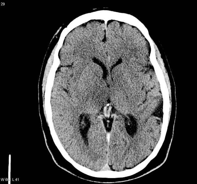
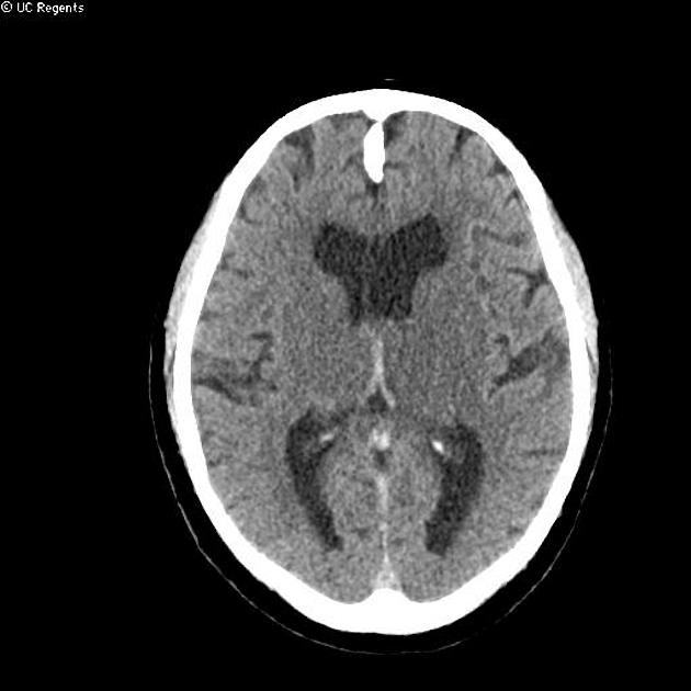
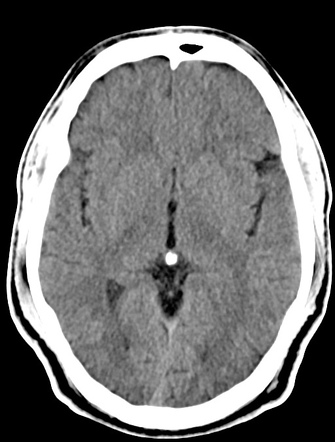
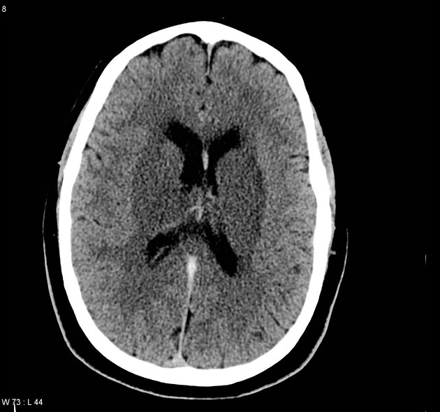
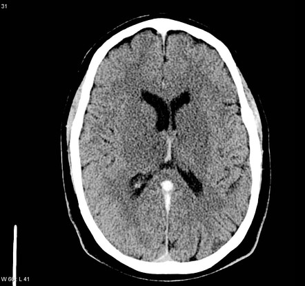
Cerebral venous infarction is an uncommon form of stroke, and is most commonly secondary to cerebral venous thrombosis and frequently manifests with hemorrhage It should be considered in infarcts (with or without hemorrhage) which do not correspond to a typical arterial territory 1. Una de las propiedades que llaman más la atención del angioma venoso radica en la discordancia entre la frecuencia de este tipo de lesiones cerebrales halladas en los estudios radiológicos y el número relativamente pequeño de personas que padecen angioma venoso. Volviendo al mundo real el caso que hoy mostramos en archivo neuroimagen es la resonancia magnética de un angioma venoso o anomalía del desarrollo venoso con su aspecto típico de cabeza de Medusa Me permito recomendar un conciso pero interesante apartado dedicado al angioma venoso de la página Angioma Alliance.
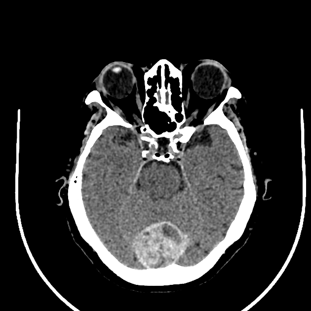
El angioma senil también se denomina "en guinda", es de color rojo brillante y suele presentarse en el tronco más que en otra partes del cuerpo Es frecuente en personas mayores de 45 años 2. Cerebral cavernous venous malformations, commonly known as cavernous haemangioma or cavernoma , are common cerebral vascular malformations , usually with characteristic appearances on MRI Cavernous malformations are found throughout the body This article focuses on cerebral cavernous venous malformations For a general discussion and links to cavernomas in other locations, please refer to the general article on cavernous venous malformation. Infratentorial Pial Angioma Of these 10 patients, in 7 (11%), there was evidence of a pial angioma involving infratentorial structures Three (43%) had involvement of both supra and infratentorial structures, and 4 (57%) had an angioma isolated to the brain stem and cerebellum.
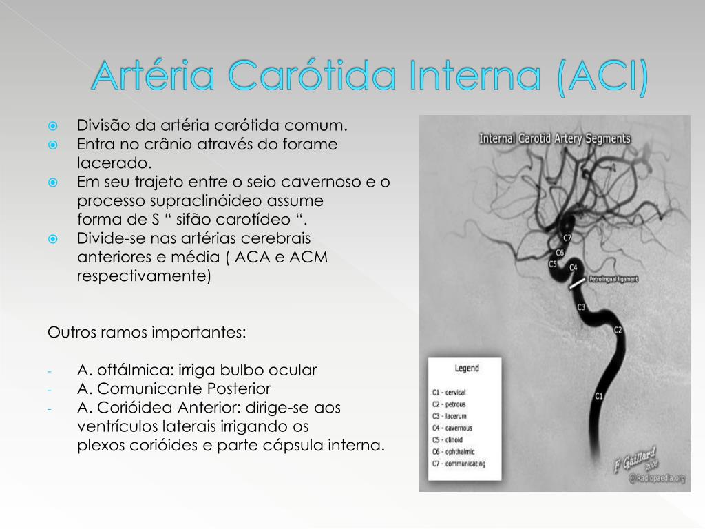
Cerebral cavernoma is a vascular malformation seen in brain and spinal cord Most of the lesions are asymptomatic These lesions are associated with epilepsy Hemorrhage can be a presenting feature It can be solitary or multiple Multiple lesions (>5 in number with a positive family history) can be seen in familial cerebral cavernous malformation. Círculo de Willis en angiografía cerebral Prof Frank Gaillard, Radiopaediaorg, rID Si bien la irrigación del encéfalo viene dada por numerosas ramas arteriales antes mencionadas, es importante acotar que varias de ellas se comunican en la porción central e inferior del cerebro, formando un sistema conocido como polígono de. Thromboembolism Superior mesenteric artery (SMA) embolus associated with cardiovascular problems (, Fig 1) is the most common cause of acute mesenteric ischemia, accounting for approximately 50% of cases (, 1)Thrombosis in the SMA or superior mesenteric vein (SMV) is a less common cause.
Las malformaciones vasculares cerebrales Se clasifican en Malformaciones arteriovenosas (MAV) , Angioma cavernoso o cavernoma, Angioma venoso , Telangiectasias Las MAV son malformaciones congénitas consistentes en ovillos vasculares con vasos aferentes arteriales y venas de drenaje, ambos displásicos, con fístulas arteriovenosas y sin intervenir la fase capilar. El abordaje inicial del paciente comprende medidas de resucitación intensivas junto con acciones dirigidas a filiar el origen del sangrado La venografía es la prueba gold estándar 10, no obstante en la actualidad la preferencia por la TAC abdominal con contraste es indiscutible, permitiendo diferenciar el origen arterial o venoso del. A developmental venous anomaly (DVA) is an unusual or irregular arrangement of small veins that may look like the spokes of a wheel The veins drain into a larger central vein DVAs are benign (not cancerous) DVAs also may be called venous angiomas or benign.
Jun 4, 17 Middle cerebral artery variants diagrams Radiology Case Radiopaediaorg.
Developmental Venous Anomaly Radiology Reference Article Radiopaedia Org

Cerebral Cavernous Venous Malformation Zabramski Type 2 Radiology Case Radiopaedia Org
Cavernoma Temporal Lobe Radiology Case Radiopaedia Org
Angioma Venoso Cerebral Radiopaedia のギャラリー
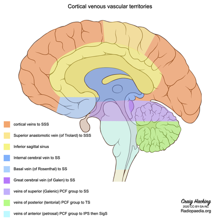
Cerebral Veins Radiology Reference Article Radiopaedia Org
Cerebral Venous Infarct Radiology Case Radiopaedia Org
Http Www Hgf Ce Gov Br Index Php Downloads Category 115 Radiologia E Diagnostico Por Imagem Download 145 3ahemangioblastoma Cerebral Relato De Caso E Revisao Da Literatura

Developmental Venous Anomaly Radiology Reference Article Radiopaedia Org
Radiology Quiz Radiopaedia Org

Developmental Venous Anomaly Radiology Reference Article Radiopaedia Org
Internal Cerebral Vein Thrombosis Radiology Case Radiopaedia Org

Thrombosed Developmental Venous Anomaly Associated With Cerebral Venous Infarct
Cerebral Cavernous Venous Malformation Radiology Reference Article Radiopaedia Org
Cavernous Haemangioma Of The Cerebellar Falx Radiology Case Radiopaedia Org

Developmental Venous Anomaly Radiology Reference Article Radiopaedia Org

Cerebral Cavernous Venous Malformation Radiology Reference Article Radiopaedia Org

Developmental Venous Anomaly Radiology Reference Article Radiopaedia Org

Signos De Neuro

Developmental Venous Anomaly Radiology Reference Article Radiopaedia Org
1

Orbital Cavernous Venous Malformation Radiology Reference Article Radiopaedia Org
Cavernoma With Bleed Midbrain Radiology Case Radiopaedia Org
Cerebral Vascular Malformations Radiology Reference Article Radiopaedia Org

Angioma Venoso Frontal P For More Information On The Advantages And Disadvantages Of Being A Real Estate Agent Please Visit Our Website Right Now P Insuficiencia Venosa Grave Icd 10 Mg
Www Neurocirugiachile Org Pdfrevista V45 N3 19 Chacon P250 V45n3 19 Pdf
1
Pontine Cavernous Hemangioma Radiology Case Radiopaedia Org
Radiology Quiz Radiopaedia Orgviewing Playlist Frcr Practise Cases Radiopaedia Org
Aqueductal Web Radiology Case Radiopaedia Org

Cerebral Cavernous Venous Malformation Radiology Reference Article Radiopaedia Org

Cerebral Venous System Radiology Reference Article Radiopaedia Org
Angiogram Of Developmental Venous Anomaly Dva Radiology Case Radiopaedia Org
Www Neurocirugiachile Org Pdfrevista V45 N3 19 Chacon P250 V45n3 19 Pdf
Deep Cerebral Vein Thrombosis Radiology Reference Article Radiopaedia Org

Venous Angioma Radiology Case Radiopaedia Org
Http Contenido Acronline Org Publicaciones Rcr Rcr28 1 08 Asimetrica Pdf

Developmental Venous Anomaly Radiology Reference Article Radiopaedia Org

Cerebral Cavernous Venous Malformation Radiology Reference Article Radiopaedia Org
Calcifying Pseudoneoplasm Of Neuraxis Radiology Case Radiopaedia Org
Http igital Dgse Uaa Mx 8080 Xmlui Bitstream Handle 1652 4350 Pdf Sequence 3 Isallowed Y
Agressive Hemangioma Radiology Case Radiopaedia Org
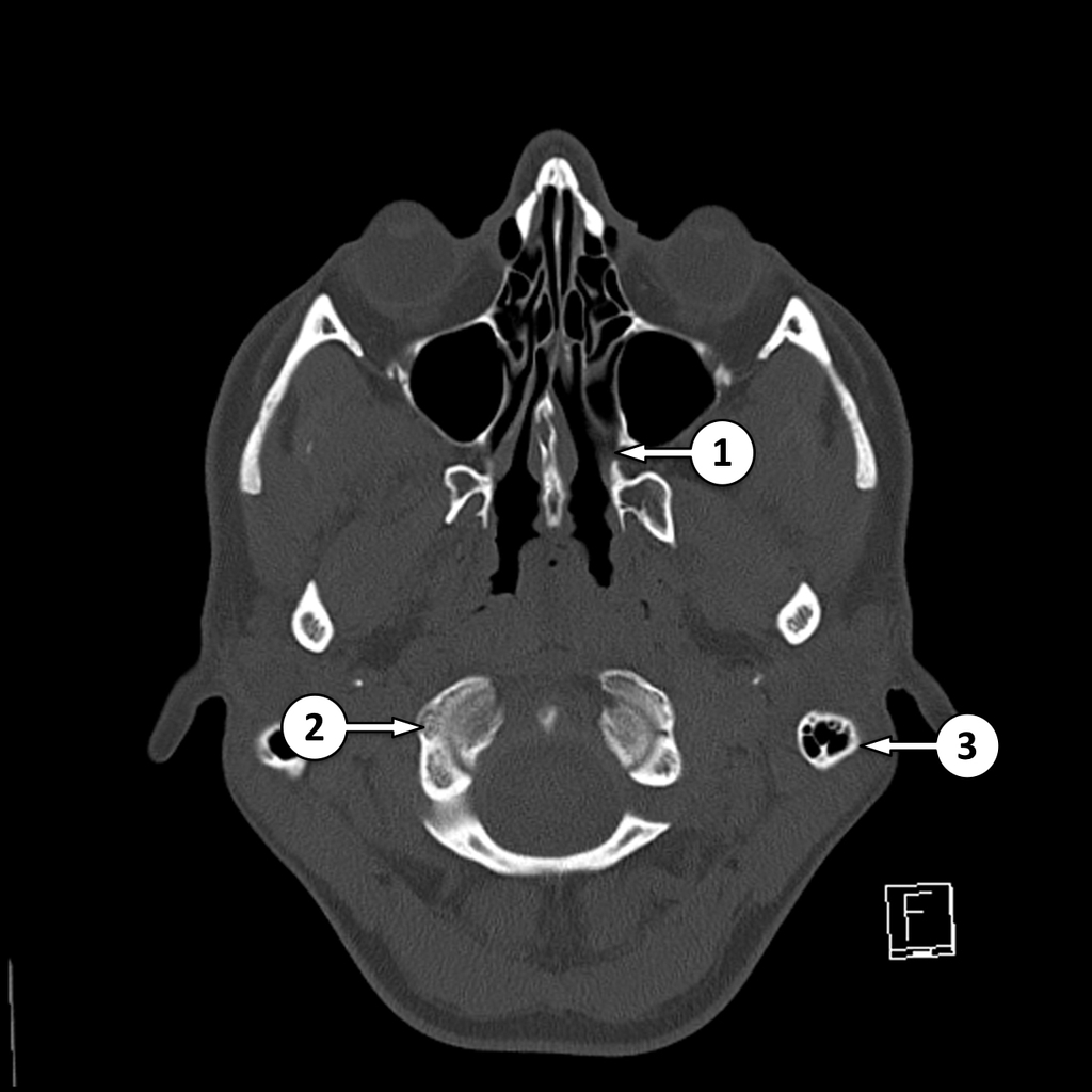
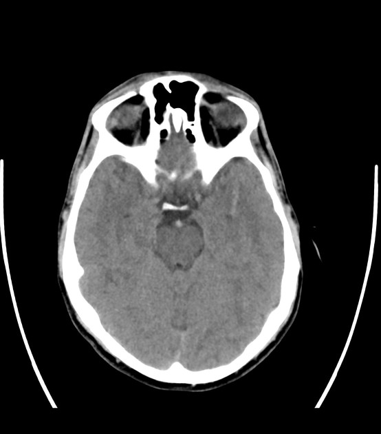
Ct Head Bone Window Axial Skull Base Labeling Questions Radiology Case Radiopaedia Org

Developmental Venous Anomaly Radiology Reference Article Radiopaedia Org

Cerebral Cavernous Venous Malformation Radiology Reference Article Radiopaedia Org
Brainstem Cavernoma Radiology Case Radiopaedia Org
Developmental Venous Anomaly Radiology Case Radiopaedia Org

Posterior Reversible Encephalopathy Syndrome Radiology Case Radiopaedia Org Radiology Brain Images Mri Brain
Malformaciones Vasculares Cerebrales Las Claves Diagnosticas Que El Radiologo Debe Conocer Pdf Descargar Libre

Signos De Neuro

Skeletal Muscle Hemangioma Radiology Case Radiopaedia Org

Radiation Induced Cerebral Vasculopathy Radiology Case Radiopaedia Org

Pin On Mri
Cerebral Vascular Malformations Radiology Reference Article Radiopaedia Org
Developmental Venous Anomaly Radiology Reference Article Radiopaedia Org
Tectal Plate Cavernoma Radiology Case Radiopaedia Org

Developmental Venous Anomaly Radiology Case Radiopaedia Org

Dandy Walker Malformation Radiology Case Radiopaedia Org Radiology Pediatric Radiology Radiology Imaging
Neurovascular Compression Of Trigeminal Nerve Radiology Case Radiopaedia Org

Sinais Em Neurorradiologia Parte 1
Q Tbn And9gcrtnumleh Ziyvv27e6dd Tulkz42yc3nuzpeie9uv57bemugdf Usqp Cau

Thalamic Cavernoma Radiology Case Radiopaedia Org
Cerebral Cavernous Venous Malformation Radiology Reference Article Radiopaedia Org
Bilateral Internal Cerebral Vein Thrombosis Radiology Case Radiopaedia Org
Giant Cerebral Cavernoma Radiology Case Radiopaedia Org

Developmental Venous Anomaly Radiology Case Radiopaedia Org

Developmental Venous Anomaly Radiology Reference Article Radiopaedia Org

Seudotumor Cerebral Radiopedia Curva De Autorregulacion Cerebral Hipertension Icd 9

Petroclinoid Ligaments Calcifications Radiology Case Radiopaedia Org Radiology Radiology Imaging Pediatric Radiology

Deep Cerebral Vein Thrombosis Radiology Case Radiopaedia Org
Cerebellar Cavernoma Radiology Case Radiopaedia Org

Cavernous Hemangioma Brain Radiology Case Radiopaedia Org

Developmental Venous Anomaly Cerebellum Radiology Case Radiopaedia Org
Cavernous Venous Malformation Radiology Reference Article Radiopaedia Org

Cerebral Cavernous Venous Malformation Radiology Reference Article Radiopaedia Org

Malformaciones Vasculares Cerebrales Las Claves Diagnosticas Que El Radiologo Debe Conocer Pdf Descargar Libre

Cerebral Vascular Territories Illustration Radiology Case Radiopaedia Org

Pdf Brain Asymmetry Diagnosis Approach
Cerebral Venous Angioma Radiology Case Radiopaedia Org
Bilateral Internal Cerebral Vein Thrombosis Radiology Case Radiopaedia Org
Developmental Venous Anomaly Radiology Case Radiopaedia Org

Cavernous Hemangioma Of The Cerebellar Falx Radiology Case Radiopaedia Org
Internal Cerebral Vein Thrombosis Radiology Case Radiopaedia Org
Q Tbn And9gcqesf3d6zgi8aoglhx5xcfx Wdeba5 Yzk4fa Pppc Usqp Cau
Cavernoma Pons Radiology Case Radiopaedia Org

Cerebral Venous Thrombosis Radiology Reference Article Radiopaedia Org
La Neuroimagen 01 01 17 02 01 17

Nb0vargcutasrm
Cerebral Venous Thrombosis Radiology Reference Article Radiopaedia Org
Developmental Venous Anomaly Radiology Case Radiopaedia Org

Pontine Capillary Telangiectasia Radiology Case Radiopaedia Org
Http Www Webcir Org Revistavirtual Articulos 15 Marzo Espana Cavernomas Ing Pdf

Developmental Venous Anomaly Radiology Reference Article Radiopaedia Org
Ppt Acidente Vascular Cerebral Powerpoint Presentation Free Download Id

Cerebral Venous Infarction Radiology Reference Article Radiopaedia Org

Calameo Manual De Anatomia Radiologica Del Distema Nervioso Central
Cerebral Venous Hemorrhagic Infarct Cortical Vein Thrombosis Radiology Case Radiopaedia Org
Developmental Venous Anomaly Radiology Reference Article Radiopaedia Org

Cerebral Venous Thrombosis Radiology Reference Article Radiopaedia Org
Angioma Venoso Radiopaedia Asimetria Fosa Yugular Medicina Para El Dolor Muscular En Pakistan

Pin On Snc
Internal Cerebral Vein Thrombosis Radiology Case Radiopaedia Org
Radiology Quiz 5217 Radiopaedia Org

Orbital Lymphatic Malformation Radiology Case Radiopaedia Org
Splenic Vein Aneurysm Radiology Case Radiopaedia Org
Developmental Venous Anomaly Radiology Case Radiopaedia Org


